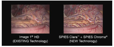
Karl Storz Endoscopy-America Inc. has announced that its Image 1 Spies Visualization Enhancement Tools system was recognized as a 2014 Innovation of the Year by the Society of Laparoendoscopic Surgeons (SLS).The new Image 1 Spies system represents the company’s latest solution for giving surgeons superior views of challenging anatomical areas during complex surgeries, ranging from skull base and orthopedic to gynecologic, urologic and thoracic.
Combined with new full-HD three-chip camera heads, Image 1 Spies includes the Clara and Chroma apps that activate proprietary image enhancement algorithms. Spies Clara automatically identifies and brightens dark areas of an image — without lag time. This addresses common problems encountered when some anatomical areas are illuminated better than others, requiring overall brightness to be increased or the scope to be moved closer to visualize darker areas. Increasing the brightness level, however, prompts tissue in the foreground to reflect light and cause glare. Moving the scope closer narrows the visible area, making it difficult to manipulate instruments and can result in mucus or blood obscuring the lens. Now, by using Spies Clara, dark areas are adjusted in real time. This avoids overexposure and reflections, providing a clear view of bright and dark regions.
Additionally, surgical interventions can be made more efficient if clear differentiation of key tissue areas is possible. Spies Chroma helps accomplish this by intensifying color contrast levels, enabling surgeons to more easily discern blood vessels and other critical anatomy. Clearly visible structure surfaces are emphasized while retaining the natural color perception of the image.